FULL TEXT OF SLIDES, Below
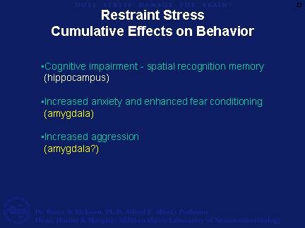
25. What happens to the behavior of the animals after chronic stress? Repeated restraint stress has a number of effects on behavior after 21d or longer. These include cognitive impairment for spatial recognition memory, increased anxiety in an open field and increased fear conditioning and increase aggression towards animals in the same cage. The cognitive impairment is likely to be related to the structural changes in the hippocampus, whereas the anxiety, fear and aggression may be due to changes in the amygdala.
A neural correlate of the increase anxiety, fear and aggression is the hypertrophy of neurons in the amygdala discovered recently by Sumantra Chattarji and colleagues in Bangalore, India, and reported in another lecture in this series.
26. An animal model of chronic psychosocial stress has been very influential and informative in showing that the hippocampal structural plasticity after chronic restraint stress can be generalized to another species and to a stressful situation that has consequences that are reminiscent of depressive illness. Tree shrews (Tupia belangeri) are solitary animals and placing an intruder to live next to a proven dominant results in considerable psychosocial stress to the intruder in terms of threats and scuffles.
Housing the intruder next to the dominant for 28d and allowing a 1h period each day when the cage door between them is open results in considerable physiological stress. Body weight declines and cortisol and catecholamines levels in the urine are increased, compared to cage controls not exposed to a dominant.
27. Repeated psychosocial stress for 28d causes remodeling of dendrites of CA3 neurons, very much like that found after repeated restraint stress and also that seen in dominant and subordinate rats in the VBS studies described earlier. Unfortunately, we do not know what happens to the dendritic remodeling in the dominant tree shrews! The remodeling of CA3 dendrites can be prevented by daily treatment of intruder tree shrews with phenytoin (Magarinos et al., 1996).
As also shown in this slide, chronic psychosocial stress also causes reduced neurogenesis in the dentate gyrus (Gould et al., 1997).
The effects of psychosocial stress to suppress neurogenesis can be prevented by daily treatment with the antidepressant, tianeptine. Other antidepressants have been reported to increase neurogenesis in the dentate gyrus, but so far the recent study by Czeh et.al. (Czeh et al., 2001) Is the only one to uses an antidepressant to counteract the effects of an ongoing stress. Tree shrews show behavioral alterations - anhedonia and reduced exploratory activity - that are prevented by treatment with antidepressants. Therefore these results have some relevance to what antidepressant may be doing in depressive illness.
28. We now return to the human brain and to a further discussion of major depressive illness.
29. At the beginning of the lecture, I mentioned the magnetic resonance imaging studies which showed that hippocampal volume loss occurs in Cushing's disease and in major depressive illness. In depression, according to Yvette Sheline and coworkers at Washington University, St. Louis, this volume loss is related to duration of the depression rather than to age per se of the patients (Sheline et al., 1999). This slide shows this relationship for both time depressed and age.
Sheline and colleagues also described in their MRI images evidence for discontinuities that might represent sites of damage. Although some recent postmortem studies on brains from depressed individuals did not show neuron loss in hippocampus, this possiblity cannot be disregarded, particularly when depression lasts a long time. How might damage come about?
30. There are specific situations where damage to the hippocampus occurs.
|