FULL TEXT OF SLIDES, Below
13. One of the ways that stress hormones modulate function within the brain is by changing the structure of neurons.
14. Within the hippocampus, the input from the entorhinal cortex to the dentate gyrus is ramified by the connections between the dentate gyrus and the CA3 pyramidal neurons. One granule neuron innervates, on the average, 12 CA3 neurons; and each CA3 neuron innervates, on the average, 50 other CA3 neurons via axon collaterals as well as 25 inhibitory cells via other axon collaterals. The net result is a 600 fold amplification of excitation as well as a 300 fold amplification of inhibition that provides negative feedback control of the system. The dentate -gyrus - CA3 system is believed to play a role in the memory of sequences of events, although long-term storage of memory occurs in other brain regions.
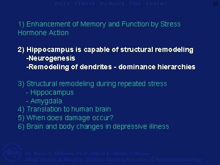
There is structural plasticity within the DG-CA3 system, in that new neurons continue to be produced in the dentate gyrus throughout adult life and CA3 pyramidal cells undergo remodeling of their dendrites.
15. The sub-granular layer of the dentate gyrus contains cells that have properties of astrocytes (they show expression of glial fibrillary acidic protein) and give rise to granule neurons (Seri et al., 2001). After BrdU administration to label DNA of dividing cells, these newly born cells appear as clusters in the inner part of the granule cell layer, where a substantial number of them will go on to differentiate into granule neurons within as little as 7 days. The new granule neurons appear to be quite excitable and capable of participating in long-term potentiation.
16. In the adult rat, 9000 new neurons are born per day and survive with a half-life of 28 days (Cameron and McKay, 2001). There are many hormonal and neurochemical modulators of neurogenesis and cell survival in the dentate gyrus. Neurogenesis in the adult dentate gyrus is enhanced by the hormone, IGF-1, and by serotonin and a number of antidepressant drugs. Estradiol accelerates cell proliferation in female rats. IGF-1 is the mediator of the ability of exercise to increase cell proliferation in the dentate gyrus. Lack of IGF-1 and insulin in diabetes has the opposite effect and decreases cell proliferation.
Neurogenesis and/or survival of newly born cells is increased by putting mice in a complex ("enriched") environment. And a form of classical conditioning that activates the hippocampus ("trace conditioning") prolongs the survival of newly-born dentate gyrus neurons.
Certain types of acute stress and many chronic stressors suppress neurogenesis or cell survival in the dentate gyrus. The mediators of these inhibitor effects include excitatory amino acids acting via NMDA receptors and endogenous opioids. We shall return to discuss stressors and neurogenesis later in the lecture.
17. The hippocampus shows another type of structural plasticity, namely, debranching of dendrites, and this can be seen in a number of naturalistic stressful situations. For example, the visible burrow system (VBS) is an apparatus with an open chamber where there is a food and water supply and several tunnels and chambers. The VBS was developed by Caroline and Robert Blanchard, University of Hawaii. Rats can be observed from above by a video camera in this apparatus. In the VBS, male rats housed with several females establish a dominance hierarchy within several days. Over the course of the next week, a few subordinate males may die and others (showing scars from bite marks) will show enlarged adrenals, low testosterone and many changes in brain chemistry. The dominant shows the fewest scars and has the highest level of testosterone but also has somewhat larger adrenal glands than cage control rats. Regarding changes in brain structure, we were surprised when we looked at the branching of dendrites within the CA3 region.
18. In the VBS, It was the dominant that had a more extensive pattern of debranching of the apical dendrites of the CA3 pyramidal neurons in the hippocampus, compared to the subordinate rats, which showed reduced branching compared to the cage controls (McKittrick et al., 2000). We will see more about this phenomenon later in conjunction with chronic stress, but what the VBS result emphasizes is that it is not adrenal size or presumed amount of physiological stress per se that determines dendritic remodeling but a complex set of other factors that modulate neuronal structure. We refer to the phenomenon as "dendritic remodeling" and we generally find that it is a reversible process. In hibernating hamsters, it occurs in a matter of hours and reverses itself just as quickly when hibernating animals are aroused from torpor.
|
