 Return to Methods List |
Methods in Neuroscience Manduca sexta Segmental Ganglia - Dissection p.1 |
| Below are photos of some of the key steps in the dissection of the Manduca larva down to the level of the ventral nerve cord. |
|
Dissection- first steps Click HERE for a 2 minute video (18.5MB) |
||
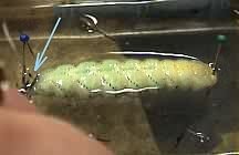 Cutting the horn off the posterior end of an anesthetized Manduca larva. |
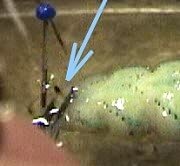 Horn shown by blue arrow. |
 Cut along entire dorsal midline. |
 All the way to the other end. |
 Using dissection pins to pull back and pin down. |
 |
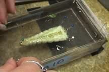 Half way done. |
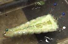 Close up view of the Manduca pinned down at the anterior end. |
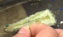 Pinning out the posterior end. |
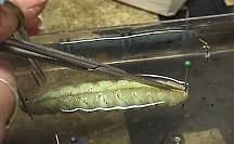
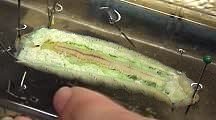 Completely pinned out. |
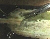
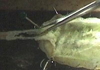
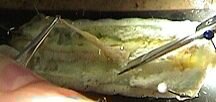 Removing the gut. |
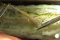 Cuts need to be made underneath to free the gut from the connecting tissue. |
 The cutting continues all the way to the the anterior end. |
 |
 |
 |
 |
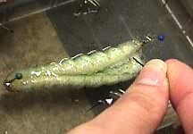
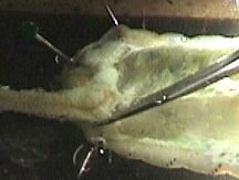
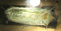 This is the end result of the first dissection. Go to the "dissection 2" page to see how the nerve cord is removed. |
| Overview | Web Lectures | Fellowships | Activities | Home | SFN | NAS | IBRO |