 Return to Methods List |
METHODS in Neuroscience Crayfish Neuromuscular Junction - DISSECTION page 2 |
|
Dissecting the First Walking Leg: Removing the shell and covering tissues to expose the muscle Click HERE for a 2 minute video of this dissection (18MB). |
|||
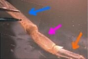
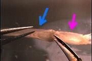
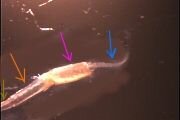
 A close-up view of the first walking leg, with the distal 3 segments (brown, orange and fuchsia arrows) glued down. The blue arrow points to the segment in which the nerve bundle is located. |
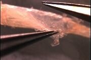
 The second segment under the dissecting microscope. The shell has been removed. |
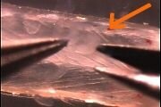
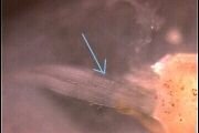
 Removing the covering tissues from the second segment. |
 |
 |
 Cutting away more of the shell on both sides of the second segment. |
 |
 |
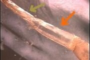 Less magnification. Much of the tissue overlying the second segment (orange arrow) muscle has been removed. |
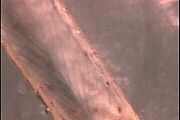
 The remaining pieces of covering tissues are gently pulled away from over the muscle. |
 Less magnification. Ready for recording. |
 Less magnification. Three of the 4 segments are visible. The segment marked by the orange arrow has been dissected. |
 The shell will be removed from the segment shown by the blue arrow. |
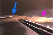 After cuts are made in the joint between the blue and fuchsia segments, the shell overlying the blue segment can be pulled off. |
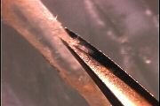
 The shell is pulled back to expose the nerve bundle and some supporting tissues (light yellow arrow). |
 The shell is almost off. |
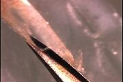
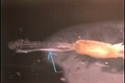 The nerve bundle (light blue arrow) and supporting tissue are free of the shell. |
 A better view of the nerve bundle (light blue arrow) held back by the forceps. |
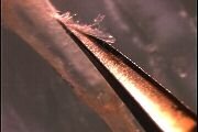
 The supporting tissue is removed. This is a close-up view of the nerve bundle as it enters the 3rd segment (the undissected segment labeled by the fuchsia arrow). |
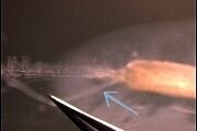
 Less magnification. All 4 segments are visible. Left most segment is distal, right most segment (nerve bundle, blue arrow) is proximal. |
| Overview | Web Lectures | Fellowships | Activities | Home | SFN | NAS | IBRO |