 Return to Methods List |
METHODS in Neuroscience Crayfish Neuromuscular Junction - DISSECTION |
|
Below are shown some of the key steps in the dissection of the crayfish neuromuscular junction, located in the the first walking leg. This particular neuromuscular junction is used because of (1) its rich repetoire of synaptic plasticity, from short to long term facilitation, (2) the accessibility of the presynaptic axon, (3) its rich repetoire of reponses to neuromodulators, (4) its simplicity.
Despite the fact that this preparation is simple, it is complicated enough to allow many types of detailed experiments to be performed. The preparation contains one excitatory axon, one inhibitory axon and a number of muscle fibers. Both axons innervate the muscle fibers and the inhibitory axon also makes presynaptic inhibitory synapses on the excitatory axon. |
|
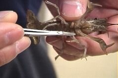
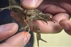
Identifying the "first walking leg" Click HERE for a short video explanation (3MB, mpg format). |
||
|
The neuromuscular junction studied is located in the first walking leg, shown by the scissors in the left photo and by the orange arrow in the right photo.
|
 |
 |
|
Mounting the first walking leg in the dissecting/recording chamber Click HERE for a short video (7MB, mpg format). |
||
| Before a recording can be made, the first walking leg must be dissected. |  The proximal portion of the leg is stuck to the blue tape so the leg can be held in position while the 3 distal segments are glued to the dish. The leg angle is adjusted so the 3 distal segments can be flat in the dish. |
 A thin streak of glue is applied to the bottom of the plastic dish. |
 The three distal segments are gently pressed down so they lie very flat. The leg needs well adhered to the dish so it does not move during the recording. |
 The job is finished. The blue tape can now be removed from the most proximal segment. This segment will later be dissected to expose the innervating nerve bundle. |
|
| The Dissecting Apparatus | |
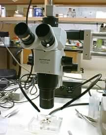 Dissecting scope with fiber optic lighting. Dissecting scope with fiber optic lighting. |
 A close-up view of the recording dish under the dissecting scope. The forceps are touching the proximal portion of leg (the part that was stuck to the blue tape, above), which will be opened to expose the nerve bundle. Click HERE for a short video giving an overview of the crayfish preparation (3MB, mpg format). |
|
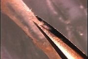
Dissecting the First Walking Leg: Removing the shell and covering tissues to expose the muscle Click HERE for a 2 minute video of this dissection (18MB). |
|||
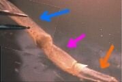
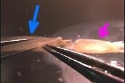
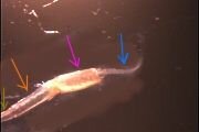
 A close-up view of the first walking leg, with the distal 3 segments (brown, orange and fuchsia arrows) glued down. The blue arrow points to the segment in which the nerve bundle is located. |
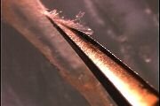
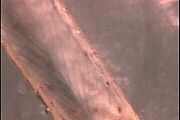
 The second segment under the dissecting microscope. The shell has been removed. |
 Removing the covering tissues from the second segment. |
 |
 |
 Cutting away more of the shell on both sides of the second segment. |
 |
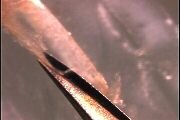 |
 Less magnification. Much of the tissue overlying the second segment (orange arrow) muscle has been removed. |
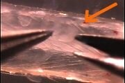 The remaining pieces of covering tissues are gently pulled away from over the muscle. |
 Less magnification. Ready for recording. |
 Less magnification. Three of the 4 segments are visible. The segment marked by the orange arrow has been dissected. |
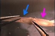 The shell will be removed from the segment shown by the blue arrow. |
 After cuts are made in the joint between the blue and fuchsia segments, the shell overlying the blue segment can be pulled off. |
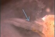
 The shell is pulled back to expose the nerve bundle and some supporting tissues (light yellow arrow). |
 The shell is almost off. |
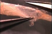
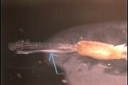 The nerve bundle (light blue arrow) and supporting tissue are free of the shell. |
 A better view of the nerve bundle (light blue arrow) held back by the forceps. |
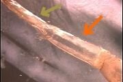
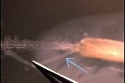
 The supporting tissue is removed. This is a close-up view of the nerve bundle as it enters the 3rd segment (the undissected segment labeled by the fuchsia arrow). |
 Less magnification. All 4 segments are visible. Left most segment is distal, right most segment (nerve bundle, blue arrow) is proximal. |
| Overview | Web Lectures | Fellowships | Activities | Home | SFN | NAS | IBRO |