 Return to Methods List |
Methods in Neuroscience Crayfish Neuromuscular Junction- RECORDING |
| Below are photos of some key steps involved in making a recording at the crayfish neuromuscular junction. Voltage- and current-clamp recordings are made with shape intracellular microelectrodes which, when filled with 3M KCl have resistances of 10-20 MOhm. |
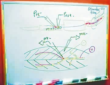
| Diagram of the recording configuration: Two recording electrodes ("pre-" & "post-") and one stimulating suction electrode |
|
|
The recording electrode labeled "pre-" is placed intracellularly in a motor axon. As you will see below, the axon is impaled at a branch point because it is easier to impale there and the recording is more stable.
The recording electrode labeled "post-" is placed in a muscle fibre innervated by the motor axon. The light blue coil in the upper right of the diagram represents the stimulating suction electrode used to stimulate the innervating nerve bundle. |
 |
| The recording equipment | |
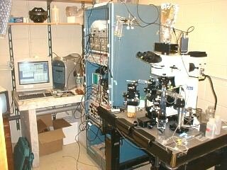 Overview of the recording set-up: computer, electronics rack, microscope |
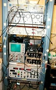 A close-up view of the electronics rack. |
 To the left of the microscope stage are the two intracellular recording electrodes. On the right is the suction electrode (green tape), used to stimulate the nerve bundle. (An additional headstage, not in use, is on the right front of the stage.) To the left of the microscope stage are the two intracellular recording electrodes. On the right is the suction electrode (green tape), used to stimulate the nerve bundle. (An additional headstage, not in use, is on the right front of the stage.) |
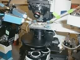 A close-up view of the microscope stage. |
| Placing the dissected preparation on the microscope stage |
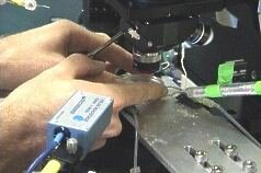 Placing the recording chamber under the scope requires some care. A recording electrode is visible in the upper left of the photo. The glass tube with the green tape contains the stimulating suction electrode. Placing the recording chamber under the scope requires some care. A recording electrode is visible in the upper left of the photo. The glass tube with the green tape contains the stimulating suction electrode. |
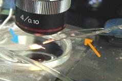 The suction electrode (orange arrow) sits above the level of the bath solution, ready to be moved into position near the exposed nerve bundle. The blue arrow shows the streak of glue used to anchor the preparation to the bottom of the dish. The preparation is not visible because of the glare of the light and glue streak. The suction electrode (orange arrow) sits above the level of the bath solution, ready to be moved into position near the exposed nerve bundle. The blue arrow shows the streak of glue used to anchor the preparation to the bottom of the dish. The preparation is not visible because of the glare of the light and glue streak. |
| Impailing the axon and muscle fibre | |
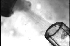
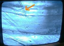 The intracellular recording electrode (orange arrow) is shown impaling the muscle fibre. The muscle fibres run from left to right across the monitor. The intracellular recording electrode (orange arrow) is shown impaling the muscle fibre. The muscle fibres run from left to right across the monitor.
Click HERE for a short video showing the electrode moving toward the neuron. You will notice the electrode vibrate when the experimenter taps on it. This is done to help the electrode impale the axon. |
|
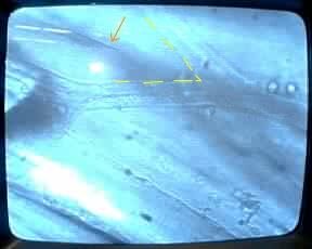 |
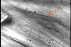 Two different views of the electrode approaching the motor axon. The electrode (orange arrow) is aiming for a branch point in the axon. In the photo at the left, the yellow dashed line outlines the "V" shape of the branch point of the axon. Two different views of the electrode approaching the motor axon. The electrode (orange arrow) is aiming for a branch point in the axon. In the photo at the left, the yellow dashed line outlines the "V" shape of the branch point of the axon. |
| Overview | Web Lectures | Fellowships | Activities | Home | SFN | NAS | IBRO |